Senior Research Projects 2017 - 2018
Seniors present to the physics department the results of their Phys 489/490: SYE Advanced Laboratory and Phys 499: Honors SYE research projects at the end of each semester. The abstracts for their research are below and photos of the student presenters follow afterwards:
Fall 2017
Extending Detection in a Lipidic Cubic Phase to Cholera and Giardia - Claire Albrecht '18
We are working on expanding the application of a detection technique to include water quality monitoring. The detection technique uses enzymatic reactions within a lyotropic liquid crystal of monolinolein. So far, we tested the conditions necessary for making the liquid crystal structure. We have also demonstrated that the enzymatic reaction required for this detection technique causes the liquid crystal to become birefringent but produced no birefringence when carried out in bulk solution.
For more information, contact Dr. Munir Pirbhai
Mechanics of Blood Flow into the Lungs of the Pre- and Post- Metamorphosed Xenopus laevis Tadpoles - Francine Nihozeko '18
In mammals, lungs become functional after birth, whereas they are functional right after metamorphosis in amphibians. The heart, on the other hand, is the first organ to develop in many vertebrate embryos. The heart is part of the circulatory system, which is necessary to transport nutrients and waste in the embryo. The cardiovascular and respiratory system are coupled. They work together to supply body tissues with fresh oxygen and to offload carbon dioxide produced during cellular respiration. Using a video frame analysis method and knowledge of fluid mechanics, this ongoing project examines blood flow into xenopus tadpoles’ lungs, to see if there are any blood flow changes associated with the lung’s function. As lungs become functional, pulmonary blood vessels expand and a non-allometric growth is expected.
For more information, contact Dr. Mike Temkin or Dr. Cristian Armendariz-Picon
High Resolution Polarization Imaging of Nacre in Iridescent Shells - Malakia Takane '18
Polarimetry is a frequently used technique in ellipsometry, astronomy, planetary science, weather radar and sometimes in computational analysis of waves. This technique is also used to measure various optical properties of materials, such as scattering birefringence and dichroism. Imaging polarimetry is a growing technique to extract information from an object that interacts strongly with polarization. Through polarimetry, the department of Physics and Astronomy at Colgate University carried out experiments that investigate the optical properties of nacre (mother of pearl) taken from several types of iridescent shells, such as bivalve pinctada gucata and gastropod abalone. Using a ×20 microscope objective, the results they obtained were of resolution ranging between 9.5 and 0.85 μm.
The goal of the project was to investigate the structure and composition of a complex biomaterial, the iridescent shell of a pearl oyster by producing high-resolution polarization images of nacre in iridescent shells. This was achieved by using a near-field scanning optical microscope (NSOM) to scan the nacre samples. A graphical programming environment, LabView, was used to control probing, collect and analyze data in order to produce high-resolution images. Using the NSOM developed by Dr. Catherine Jahncke, polarization images of higher special resolution than those obtained in the nacre polarimetry experiment carried out at Colgate University were obtained.
For more information, contact Dr. Catherine Jahncke
Spring 2018
Progress towards a point-of-care diagnostic tool involving lyotropic liquid crystals - Claire Albrecht '18
This work is geared towards the field of point-of-care diagnostics, which aims at developing rapid, reliable and cheap disease detection techniques for non-laboratory settings. It builds on the work of Vallooran et al. who have used a sequence of enzymatic reactions within lyotropic liquid crystals to detect biomarkers, viruses, bacteria, and parasites. Our work takes a step towards making their diagnostic technique more cost-effective. For example, we have replaced the polarizing microscope (which can cost hundreds of dollars) in the detection scheme with a pair of cheap, crossed film polarizers (~$2). We also show that the use of a spectrometer in the setup can limit false-positive results.
For more information, contact Dr. Munir Pirbhai
Presenter
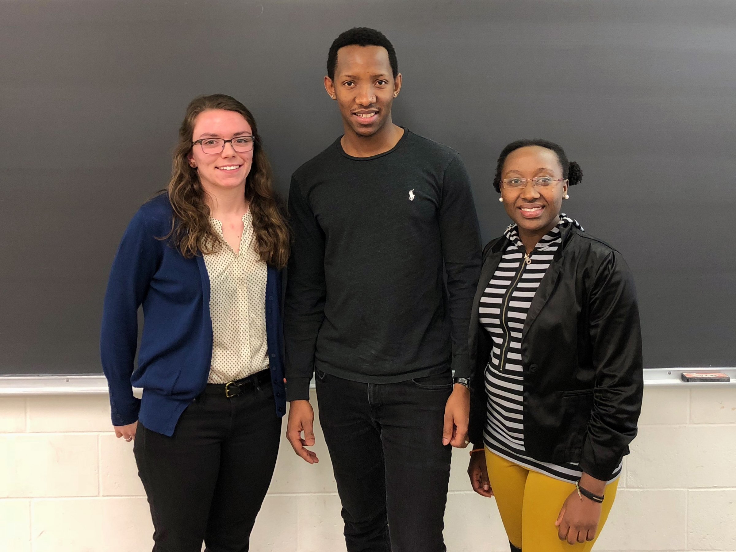
Senior presenters
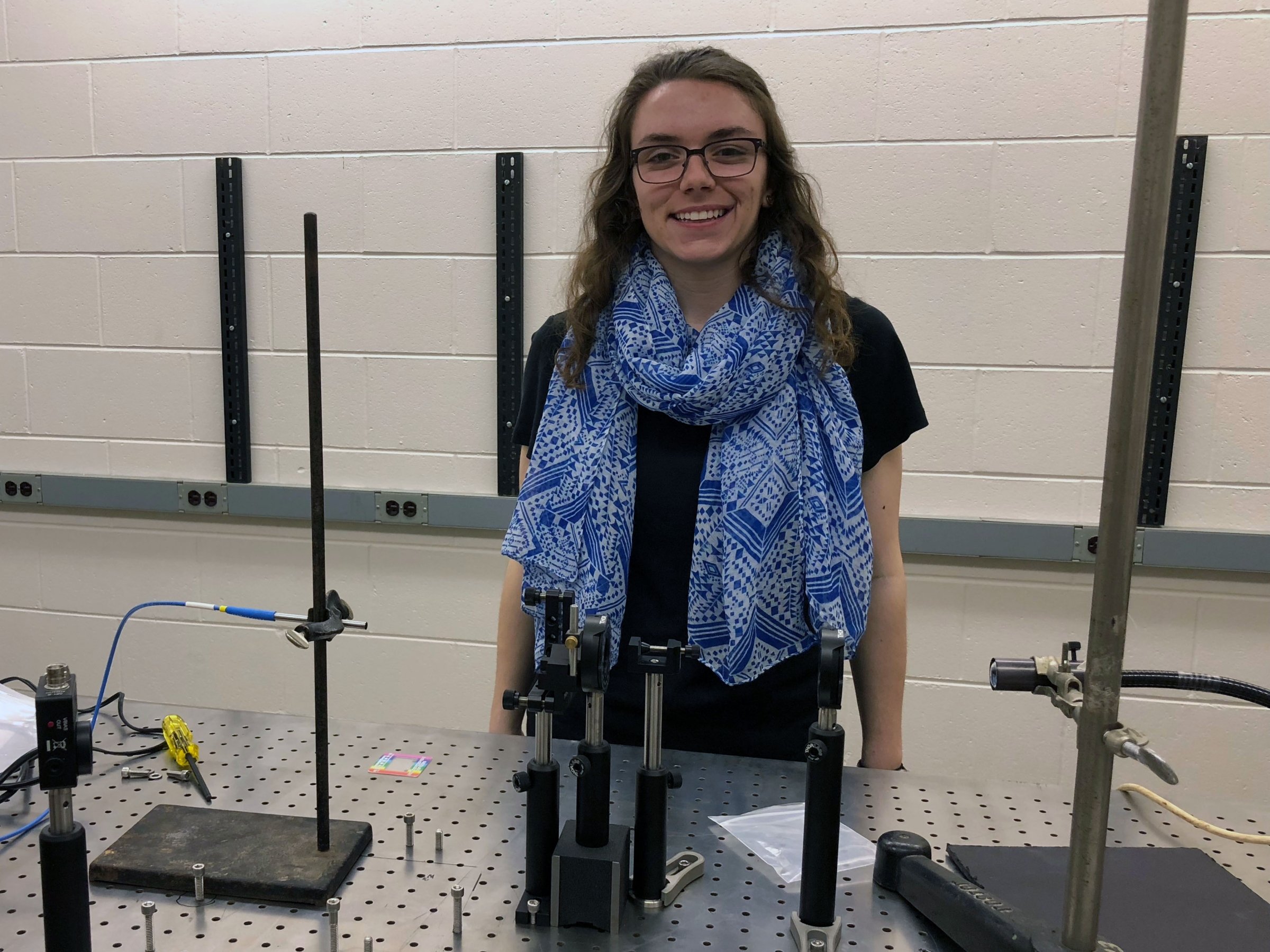
Claire Albrecht
Extending Detection in a Lipidic Cubic Phase to Cholera and Giardia
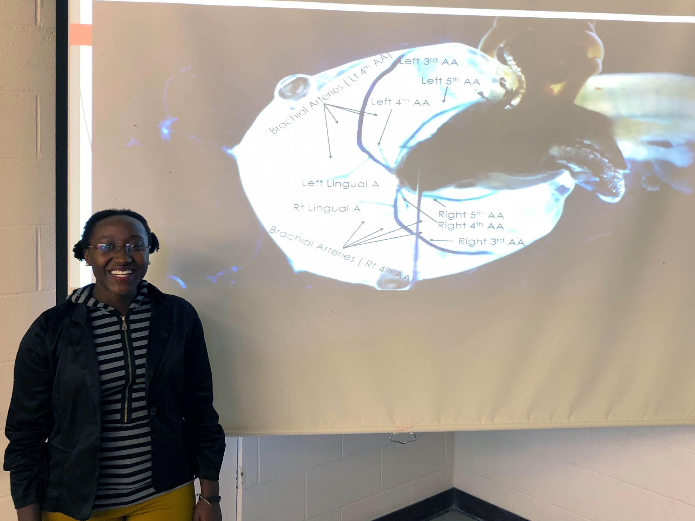
Francine Nihozeko
Mechanics of Blood Flow into the Lungs of the Pre- and Post- Metamorphosed Xenopus laevis Tadpoles
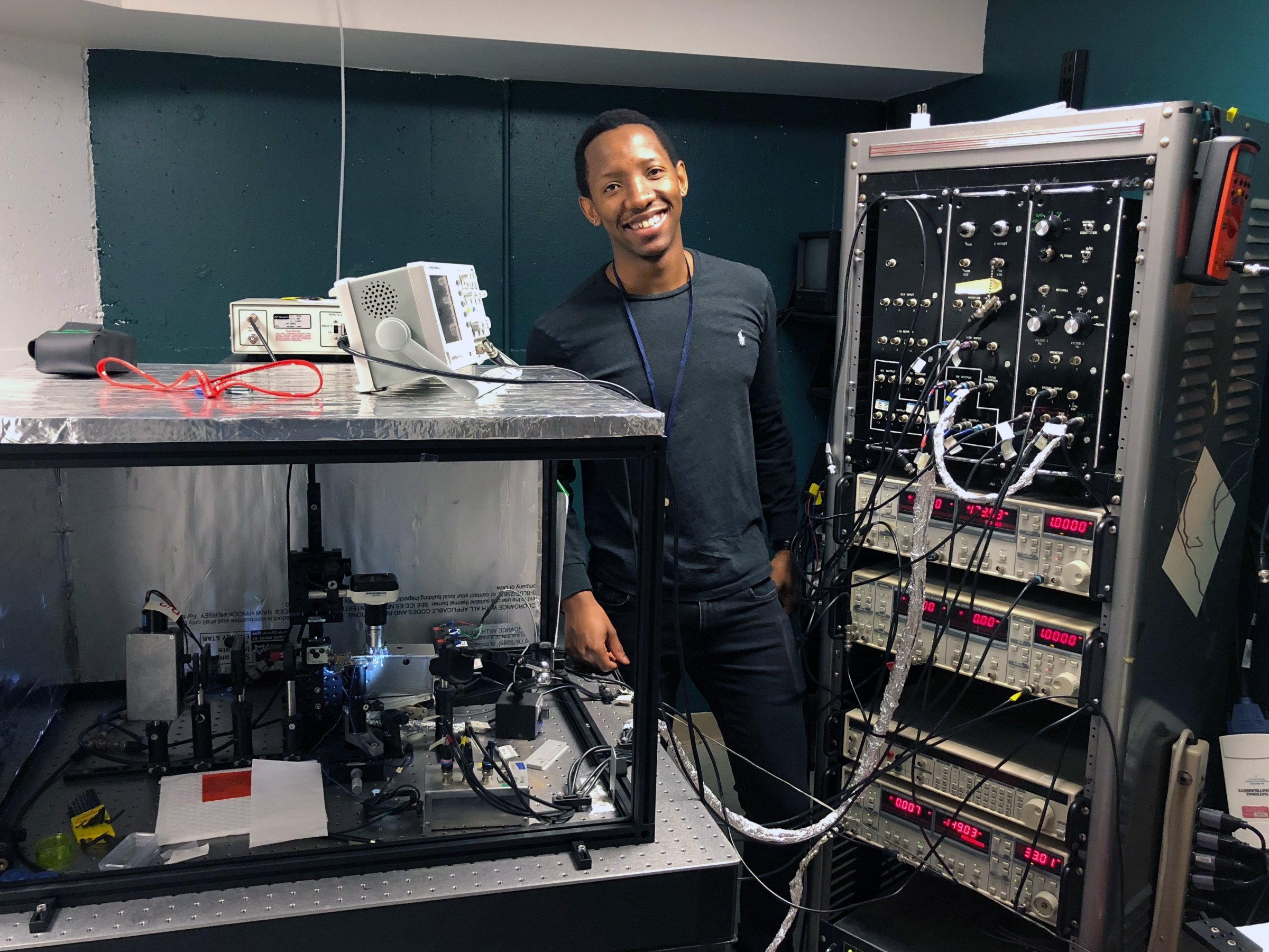
Malakia Takane
High Resolution Polarization Imaging of Nacre in Iridescent Shells